Localization studies
Ultrasound
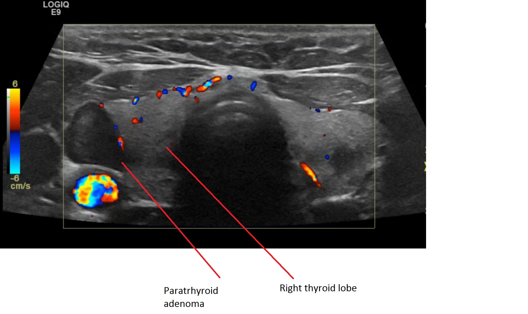
Ultrasound is the most frequently anatomic imaging modality used in the evaluation of thyroid and parathyroids. US does not use ionizing radiation and is cheaper compared to other imaging techniques but is highly user-dependant and does not enable retromanubrial or mediastinal visualization. Normal parathyroid glands are rarely visualized by ultrasonography, because of their small size and insufficient acoustic difference compared to adjacent thyroid tissue. Parathyroid adenomas, hyperplasia, and carcinomas exhibit a relatively hypoechogenic pattern, due to their compact cellularity relative to thyroid tissue. US in highly sensitive in experienced hands with sensitivity of up to 80%. If thyroid disease/ nodules identified at the time of evaluation of parathyroid, further evaluation of the thyroid should be considered prior to parathyroid surgery.
Sestamibi Scan
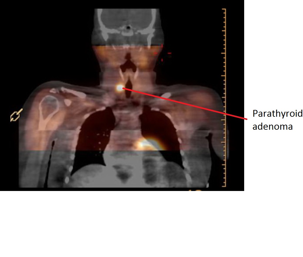
Sestamibi (Technetium-99m-methoxyisobutyl isonitrile) scan is a nuclear medicine imaging test used in identifying parathyroid adenoma. It is taken up by both thyroid and parathyroid tissue; however, it is retained by abnormal parathyroid tissue. Presence of thyroid nodules may decrease the sensitivity of this imaging technique. Addition of single photon emission computed tomography (SPECT) is shown to increase the sensitivity and specificity of parathyroid imaging allowing for better localization of the enlarged glands. Frequently, sestamibi scan is used in conjunction with some type of anatomic imaging, either ultrasound, CT, or MRI in evaluating abnormal parathyroid glands.
Computed Tomography (CT) and Magnetic Resonance Imaging (MRI)
CT scanning can provide valuable and complimentary localizing information for the identification of abnormal parathyroid glands. This technique relies on the vascularity of the parathyroid glands and their relative enhancement with contrast compared to the surrounding structures CT scanning may be useful in identifying parathyroid glands in atypical locations. MRI is another anatomic imaging modality used for detecting abnormal parathyroid glands. This method is preferred by some over CT scanning due to lack of ionizing radiation.
Parathyroid Localization Studies
|
Noninvasive |
Invasive |
|
High-resolution US Tc 99m Sestamibi scan SPECT-CT MRI CT scan |
Selective venous sampling Angiogram US guided FNA US guided Jugular Venous sampling |


